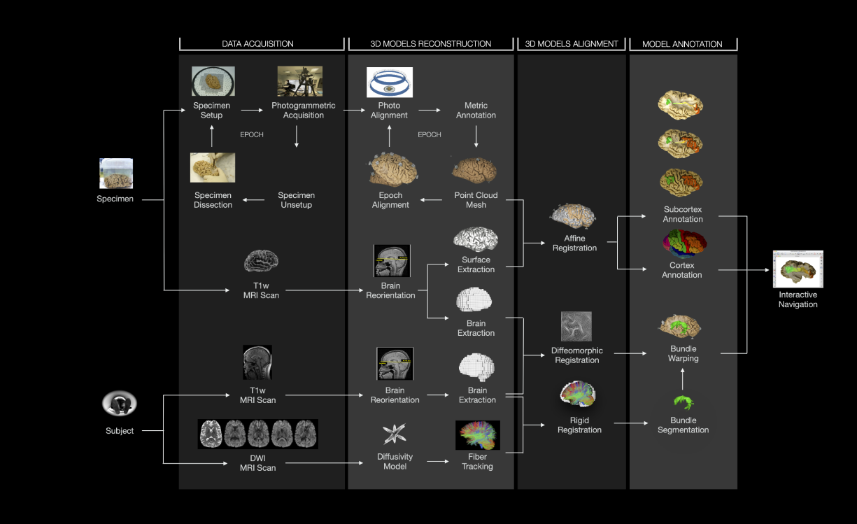
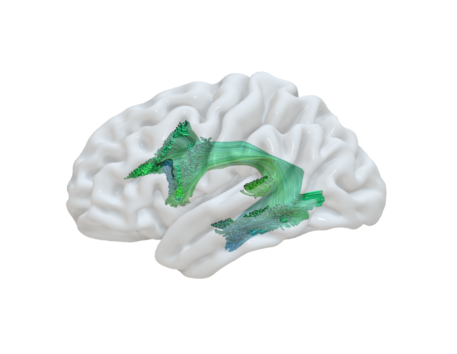
Integrated visualization of ex-vivo and in-vivo data derivatives.
The CloudCompare visualization tool is
supporting both point clouds and meshes representation

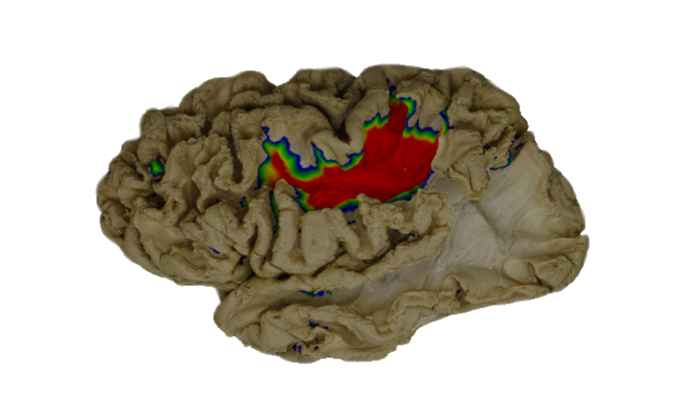
Identification and segmentation of the white matter pathways of interest on the specimen's point cloud along the dissection steps
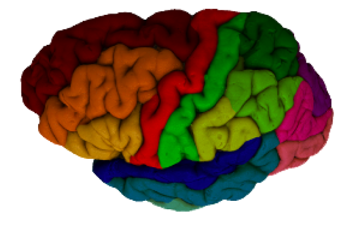
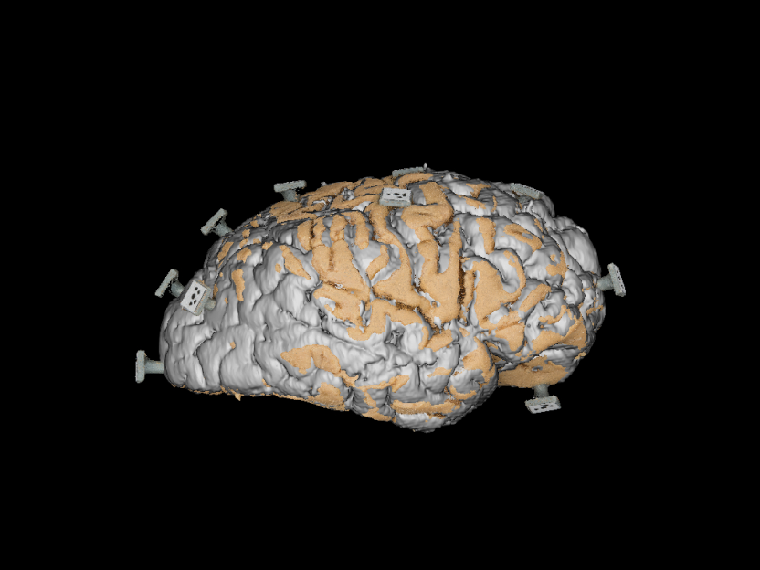
Segmentation of the gray matter on the specimen's point cloud according to gyral/sulcal macroanatomical landmarks
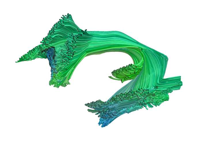
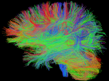
Manual segmentation of the bundle of interest from the whole brain tractography reconstructed from in-vivo diffusion MR images.
Registration of the bundle tractography from the individual MRI space to the reference space of the specimen.
Computation of linear transformation between photogrammetric and MRI models of the specimen
Computation of non linear transformation between the T1W of the specimen and individual MRI
In-vivo images alignment. Registration of the tractography from DWI space to the T1W space of the same individual.
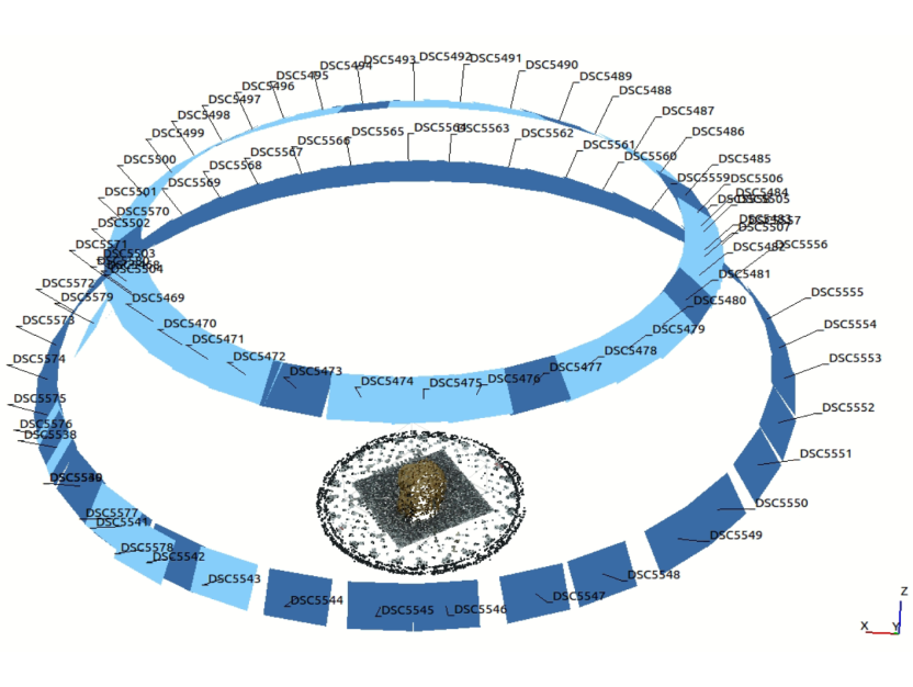
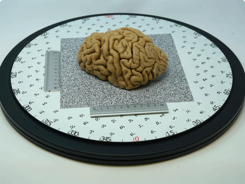
Reconstruction of reference system for the photogrammetric acquisition and annotation of metric system
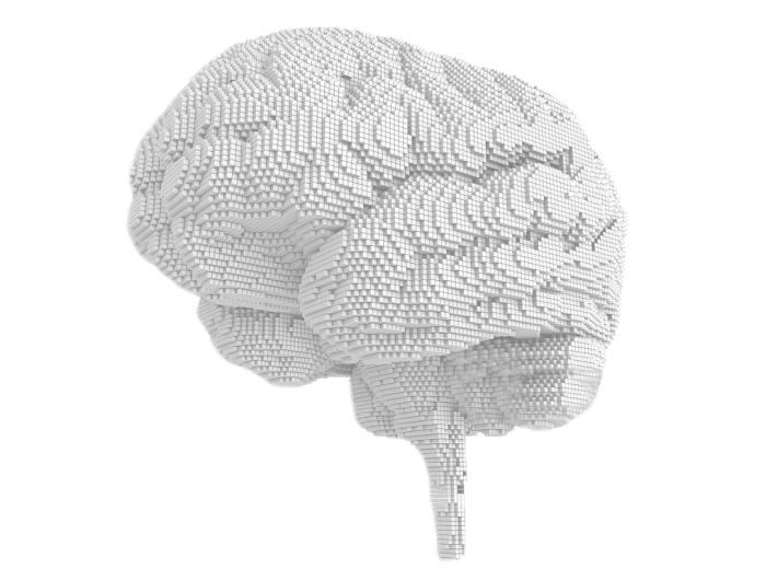
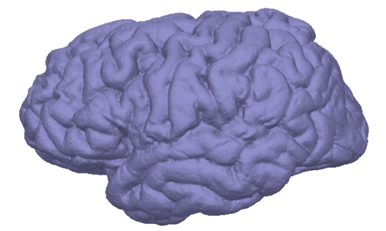
Reconstruction of the 3D model and related texture of the specimen
Rigid transformation for the alignment of two subsequent epochs
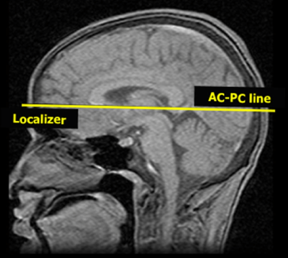
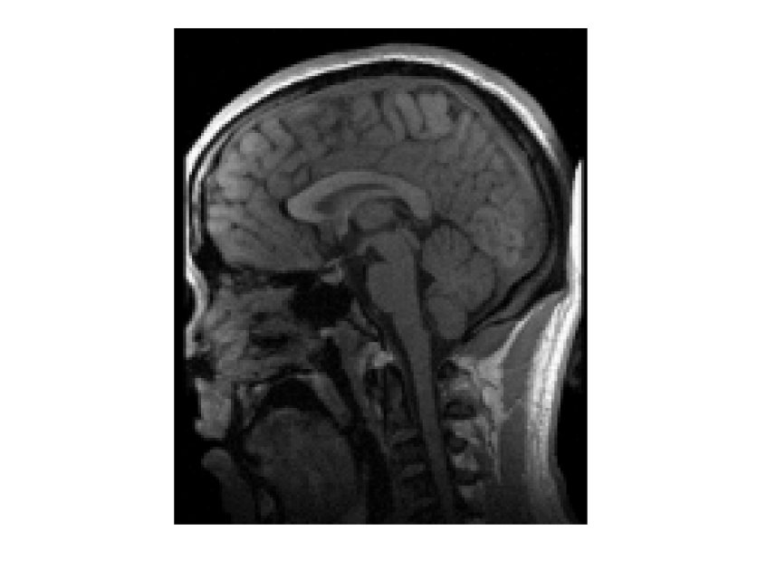
ACPC alignment of the T1 weighted structural brain image both for individual and specimen
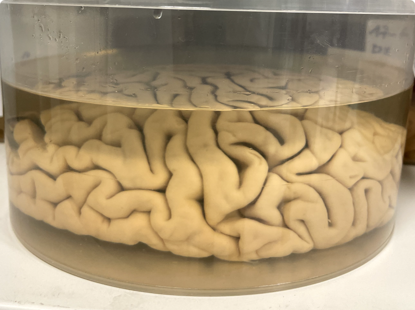
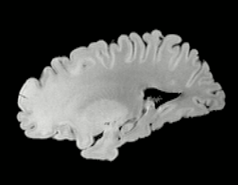
Acquisition of a T1 weighted structural image of the ex-vivo brain before the session of dissection
Skull removal to obtain brain volume only
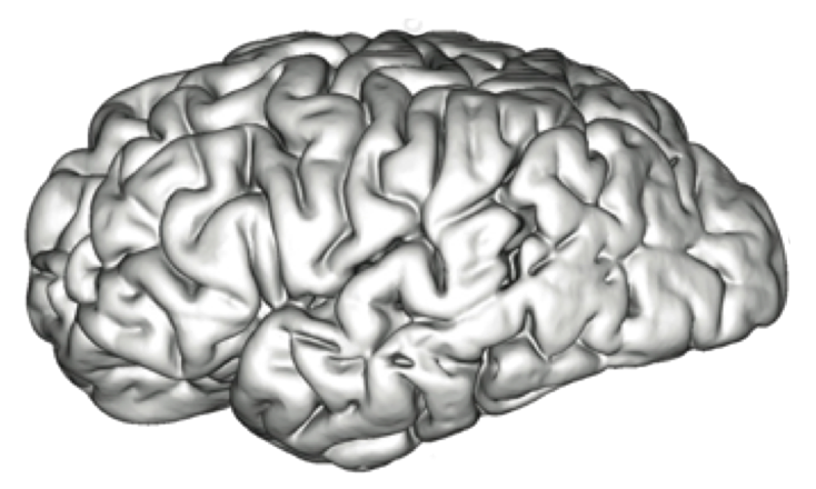
Computation of the cortical mesh from the T1 weighted MR image of the ex-vivo brain
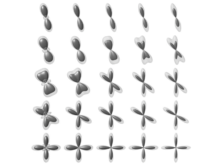
Computation of constrained spherical deconvolution model
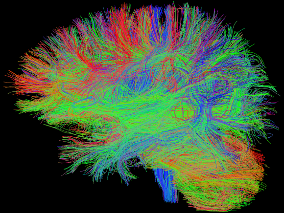
Computation of the whole brain tractogram with CSD model

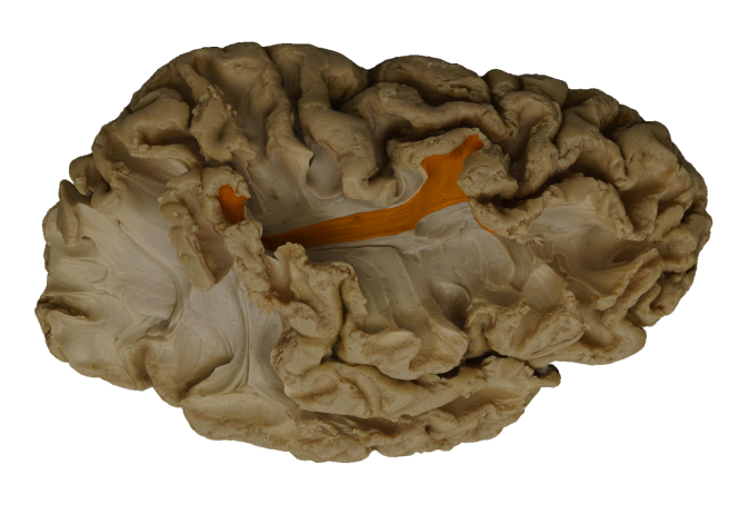
Iterative sessions of dissection of the cortical and subcortical tissues
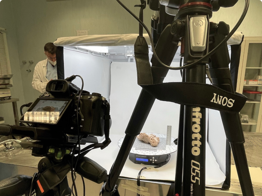
Positioning of the specimen on the referenced table at the current epoch
Photogrammetric acquisition of the current epoch of dissected specimen
Acquisition of T1 weighted structural brain image
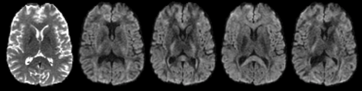
Acquisition of a sequence of Diffusion Weighted Images
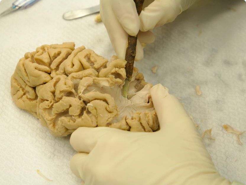

Klingler preparation of a single hemisphere after autopsy session (Sarubbo S et al., 2015)
Healthy volunteer individual for an MRI session imaging acquisition