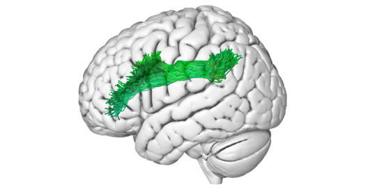
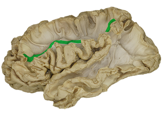
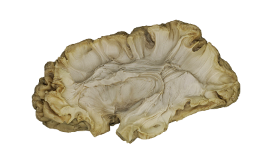
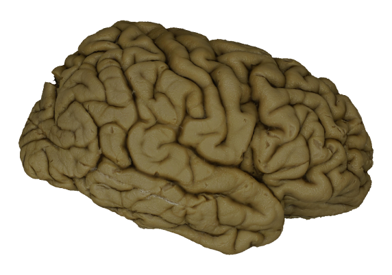
TRACTOGRAPHY
White Matter
Fiber Bundles
full_tractography 1
ANNOTATIONS
Cortical and subcortical ROIs
004
DISSECTIONS
3D photogrammetry models
008
Data

CATALOGUE

BRADIPHO
Download
Preview
Tool
Workflow
Data
About
Workflow
Download
Preview
TRACTOGRAPHY

Connection AG-STG
The bundle that connects the Angular Gyrus and the Superior Temporal Gyrus

Connection AG-SPG
The bundle that connects the Angular Gyrus and the Superior Parietal Gyrus

Connection AG-SMG
The bundle that connects the Angular Gyrus and the Supramarginal Gyrus

Connection AG-PrCG
The bundle that connects the Angular Gyrus and the Precentral Gyrus

Connection AG-PoCG
The bundle that connects the Angular Gyrus and the Postcentral Gyrus

Connection AG-MTG
The bundle that connects the Angular Gyrus and the Middle Temporal Gyrus

Connection AG-MFG
The bundle that connects the Angular Gyrus and the Middle Frontal Gyrus

Connection AG-ITG
The bundle that connects the Angular Gyrus and the Inferior Temporal Gyrus

Connection AG-IOG
The bundle that connects the Angular Gyrus and the Inferior Occipital Gyrus

LEFT
RIGHT
Line 16 sub-01_epo-01_ref-00_lateral sub-01_epo-01_ref-00_lateral sub-01_epo-01_ref-00_lateral sub-01_epo-01_ref-00_lateral sub-01_epo-01_ref-00_lateral sub-01_epo-01_ref-00_lateral sub-01_epo-01_ref-00_lateral sub-01_epo-01_ref-00_lateral sub-01_epo-01_ref-00_lateral sub-02_epo-01_ref-00_lateral sub-02_epo-01_ref-00_lateral sub-02_epo-01_ref-00_lateral sub-02_epo-01_ref-00_lateral sub-02_epo-01_ref-00_lateral sub-02_epo-01_ref-00_lateral sub-02_epo-01_ref-00_lateral sub-02_epo-01_ref-00_lateral sub-02_epo-01_ref-00_lateral

The collection of 11 bundles segmented from tractography of an healthy individual and representing the connectivity of Angular Gyrus. The bundles are registered to the specimen space (Petit L et al., 2023).

ANNOTATIONS

Cortical ROI
19 annotations delimiting the cortical gyri of the lateral and basal surface of the hemisphere are provided for each photogrammetric model and each dissection epoch.
As the dissection proceeds according to the latero-medial approach adopted, only the spared gyri (or the remaining part of them) are represented.

LEFT
RIGHT
Line 15 sub-01_epo-01_ref-00_lateral sub-01_epo-01_ref-00_lateral sub-01_epo-01_ref-00_lateral sub-01_epo-01_ref-00_lateral sub-01_epo-01_ref-00_lateral sub-01_epo-01_ref-00_lateral sub-01_epo-01_ref-00_lateral sub-01_epo-01_ref-00_lateral sub-02_epo-01_ref-00_lateral sub-02_epo-01_ref-00_lateral sub-02_epo-01_ref-00_lateral sub-02_epo-01_ref-00_lateral sub-02_epo-01_ref-00_lateral sub-02_epo-01_ref-00_lateral sub-02_epo-01_ref-00_lateral sub-01_epo-01_ref-00_lateral

The collection of the manual annotations of the main brain structures. Cortical ROIs have been segmented along the progression of the dissection on the 3D photogrammetric model of each epoch.
A similar process of annotation has been carried out detecting the portion of the main bundle pathways exposed by the subcortical dissection (Petit L et al., 2023).

Connection ROI: AG-IFG
The pathway that connects the Angular Gyrus and the Inferior Frontal Gyrus

Connection ROI: AG-MFG
The pathway that connects the Angular Gyrus and the Middle Frontal Gyrus

Connection ROI: AG-MTG
The pathway that connects the Angular Gyrus and the Middle Temporal Gyrus

Connection ROI: AG-PoCG
The pathway that connects the Angular Gyrus and the PostCentral Gyrus

Connection ROI: AG-PrCG
The pathway that connects the Angular Gyrus and the PreCentral Gyrus

Connection ROI: AG-SMG
The pathway that connects the Angular Gyrus and the Supramarginal Gyrus

Connection ROI: AG-SPG
The pathway that connects the Angular Gyrus and the Superior Parietal Gyrus

sub-02_epo-01_ref-00_lateral sub-01_epo-01_ref-00_lateral sub-02_epo-01_ref-00_lateral sub-01_epo-01_ref-00_lateral

Connection ROI: AG-STG
The pathway that connects the Angular Gyrus and the Inferior Frontal Gyrus

sub-02_epo-01_ref-00_lateral sub-01_epo-01_ref-00_lateral

Connection ROI: AG-STG
The pathway that connects the Angular Gyrus and the Inferior Frontal Gyrus

DISSECTIONS

The collection of 3D photogrammetric models of ex-vivo specimens.
Two hemispheres are available, one left and one right.
For each hemisphere a brain model has been reconstructed to capture the progression of tissue removal across the sequence of epochs for the Superior Longitudinal System (Mandonnet et al., 2018).

EPOCH 1
Lateral surface of the hemisphere

EPOCH 2
Decortication of the sulci

EPOCH 3
Exposure of the superficial layer of the posterior transverse system

EPOCH 4
Exposure of the deep layer of the posterior transverse system

EPOCH 5
Exposure of the superficial layer of the Superior Longitudinal System: fronto-parietal part (SLF)

EPOCH 6
Exposure of the deep layer of the Superior Longitudinal System: fronto-parietal part (SLF)

EPOCH 7
Exposure of the superficial layer of the Superficial Longitudinal System: fronto-temporal part (AF)

EPOCH 8
Exposure of the fronto-temporal Superior Longitudinal System (AF) stem

EPOCH 9
Removal of the operculum with the exposure of the insula

Left hemisphere not available

EPOCH 10
Removal of the insula, extreme capsule and claustrum with the exposure of the external capsule - superficial layer of the inferior longitudinal system (IFOF and UF)

EPOCH 11
Delineation of the IFOF stem

LEFT
RIGHT
Line 12 sub-01_epo-01_ref-00_lateral sub-01_epo-01_ref-00_lateral sub-01_epo-01_ref-00_lateral sub-01_epo-01_ref-00_lateral sub-01_epo-01_ref-00_lateral sub-01_epo-01_ref-00_lateral sub-01_epo-01_ref-00_lateral sub-01_epo-01_ref-00_lateral sub-01_epo-01_ref-00_lateral sub-01_epo-01_ref-00_lateral sub-01_epo-01_ref-00_lateral sub-02_epo-01_ref-00_lateral sub-02_epo-01_ref-00_lateral sub-02_epo-01_ref-00_lateral sub-02_epo-01_ref-00_lateral sub-02_epo-01_ref-00_lateral sub-02_epo-01_ref-00_lateral sub-02_epo-01_ref-00_lateral sub-02_epo-01_ref-00_lateral sub-02_epo-01_ref-00_lateral sub-02_epo-01_ref-00_lateral sub-02_epo-01_ref-00_lateral